Summary
of Embryonic Development
Unfortunately,
due to the volume of material that we must work through in this course,
embryology is one of the topics that won’t be covered in great detail. Embryology
is the study of the development of living organisms from fertilized egg, or
zygote, to birth. This field of study is
infinitely interesting and furthers the understanding of numerous congenital
conditions. During the study of organ
systems in this course, students will frequently come across embryologic terms
such as blastocyst, mesoderm, endoderm, and ectoderm. For this reason, a brief summation is
presented here.
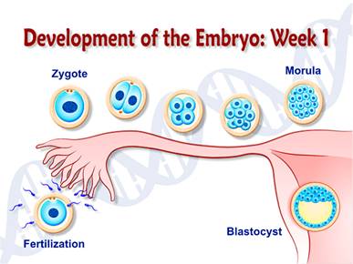
The
diagram below illustrates human development in the first week of life. Note that the fertilized egg is called a zygote.
That cell is surrounded in a protective corona. Within the corona, the zygote cells divides
multiple times. The cells then organize
themselves into the blastocyst. The blastocyst must embed itself in the
upper third of the lining (endometrium)
of the uterus if the pregnancy is to be successful.
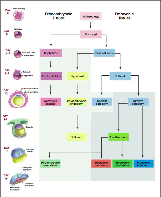
While
buried in the endometrium, the blastocyte undergoes a series of cell migrations
called gastrulation. This process begins when cells on the surface
dive inward to become one of the three germ layers. A second and then third invagination of cells
leads to development of the embryo. The
chart on the next page illustrates the terminology, timeline, and development
of the 3 germ layers: ectoderm,
mesoderm, and endoderm.
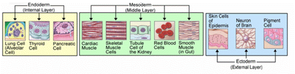
Now that we have
considered the development of the multilayered organization of the embryo, let
us consider the fate of the three germ layers.
Each germ layer is destined to produce a specific line of cells that becomes
different organ systems. The diagram
below sums up this process. (I would
like to add that endoderm forms the organs of the digestive system.)
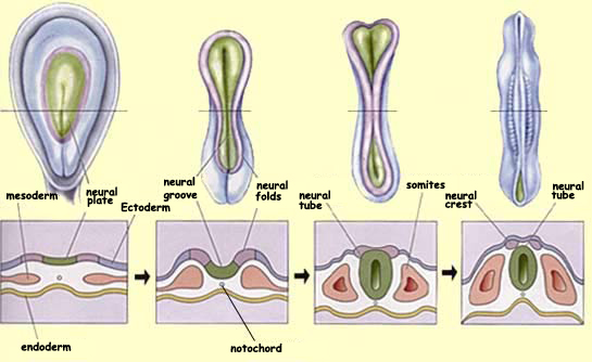
The
development of the nervous system begins with the formation of the neural plate from the ectoderm layer at
day 19. Two ridges form on the margin of
the neural plate that will become the neural
folds. The neural plate deepens to
form the neural groove by day
20. By the 22nd day, the
neural folds have formed the neural
crests which will become the cranial and spinal nerves. The neural groove closes to become the neural tube by day 26.
This
early development of the nervous system, completed by the end of the 4th
week of fetal development, is illustrated in the diagrams below.
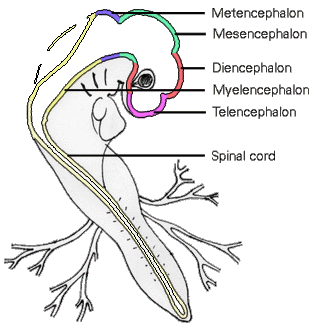
As
soon as the neural tube forms, its anterior end begins to expand and
constrictions appear. The resulting
three primary brain vesicles are, from the anterior tip back, the prosencephalon (forebrain), mesencephalon (midbrain), and rhombencephalon (hindbrain).
In week 5, the
primary vesicles will give rise to the secondary brain vesicles. The prosencephalon divides into the telencephalon and diencephalon. The mesencephalon remains undivided. The rhombencephalon divides into the metencephalon and myelencephalon. Below are
two diagrams of a five week fetus, specifically showing further development of
the nervous system.
|
|
|
The
table below sums up what the embryonic structures of the nervous system become
in adults.
|
Neural Tube |
Primary Brain Vesicles |
Secondary Brain Vesicles |
Adult Brain Structures |
|
Anterior End |
Prosencephalon (forebrain) |
Telencephalon |
Cerebrum: Cerebral hemispheres (cortex, white matter,
basal nuclei) |
|
Diencephalon |
Diencephalon (Thalamus,
hypothalamus, epithalamus) |
||
|
Mesencephalon (midbrain) |
Mesencephalon |
Brain stem: Midbrain |
|
|
Rhombencephalon (hindbrain) |
Metancephalon |
Brain stem: pons |
|
|
Cerebellum |
|||
|
Myelencephalon |
Brain stem: medulla oblongata |
||
|
Posterior End |
|
|
Spinal cord |
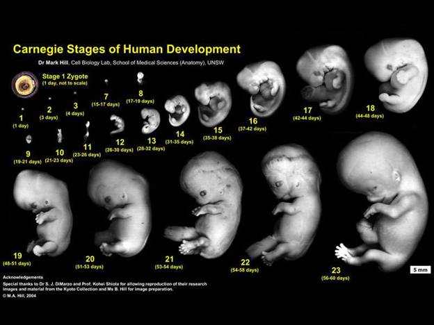
This series of sonogram
pictures shows fetal development from day 1 to day 60 (roughly through the
first 2 months of pregnancy).